*RefereeBio

FAQ

Frequently asked questions about private manuscript review.

RefereeBio is a pre-submission preparation tool. These answers summarize the most important privacy, affiliation, and responsible-use limits.

WHAT REFEREE ANALYZES & PREPARES

Analysis, reviewer reasoning, and a practical revision plan.

Each report assesses the manuscript against the expectations of its journal context, then turns the most important risks into clear next steps.

Novelty & conceptual depth Mechanistic evidence Statistical rigor Figure clarity Scope & journal fit Desk-triage risk Reviewer perspectives Statistical audit Section analysis Ranked fixes Prior Art & Novelty Risk Point-by-point rebuttal generation

Is my manuscript private?

Your manuscript is used to generate your private report, saved history, exports, and requested editing or revision check features. RefereeBio does not publish your uploads, submit them to journals, or sell manuscript content.

Does Referee train on my manuscript?

RefereeBio does not intentionally use private manuscripts to train public AI models. Manuscript text may be sent to configured AI providers to generate requested output, subject to provider terms and data-processing settings.

What does Referee not do?

Referee does not make publication decisions, contact editors, submit manuscripts, certify statistics, replace collaborators or compliance review, or promise that a revision will change any journal outcome.

Is Referee affiliated with journals?

No. Journal and publisher names are used descriptively for submission-planning context only. RefereeBio is independent and is not affiliated with, endorsed by, sponsored by, certified by, or operated by any journal, publisher, editor, society, or indexing service.

Can I upload patient data or PHI?

No, not by default. Do not upload protected health information, patient-identifying data, HIPAA-covered data, FERPA-covered records, export-controlled data, classified data, or other specially regulated content unless RefereeBio signs a separate written agreement covering that data.

Can I delete uploaded manuscript files?

Yes. Uploaded manuscript files can be deleted while generated reports remain in your account history. Some operational records may remain temporarily in backups, logs, payment records, provider systems, or security records where required.

UNDERSTANDING YOUR RESULTS

What does my score mean?

Referee reports several different signals. They answer different questions and should not be read as interchangeable grades or publication probabilities. Read the methodology for a full explanation of each dimension.

Overall manuscript quality score

This is a journal-independent assessment of the manuscript in its current form. Each dimension is built from specific, checkable evidence found directly in the manuscript's own text — not a single holistic impression — so the score reflects concrete, identifiable features of the manuscript rather than an overall vibe.

The assessment first identifies what kind of study this is — a mechanistic experiment, a genetic or chemical screen, a computational method, a clinical trial, a structural study, and so on — and evaluates the relevant dimensions against evidence standards appropriate to that study type. A screening paper isn't penalized for lacking a rescue experiment it was never designed to run; a computational method isn't judged by wet-lab standards. Many manuscripts blend more than one study type, and the assessment accounts for that blend rather than forcing a single category.

The ten dimensions carry different weight, with statistical confidence, claim–evidence alignment, and methodological robustness weighted most heavily; figure strength contributes when figures are available. Scores are calibrated against real manuscripts so the numbers mean something comparable across very different papers: a genuinely average, competent manuscript should land in the mid-70s, work strong enough for a well-regarded but non-flagship journal typically lands in the low-to-mid 80s, and only work that is exceptional across nearly every evaluated dimension reaches the 90s. It is not an acceptance probability, journal percentile, or editorial decision.

The quality assessment reads the complete extracted manuscript, including later results, methods, legends, supplements, limitations, and statistical details. An identical normalized manuscript reuses the same versioned assessment regardless of the journal selected; journal choice changes the reviewer commentary and journal-fit analysis, not this score.

Individual manuscript scores

Novelty
How unexpected, original, or field-advancing the central contribution appears.
Conceptual depth
How clearly the work develops a meaningful model, framework, or testable explanation.
Mechanistic depth
How directly the evidence establishes mechanism rather than association alone.
Technical strength
The quality of execution, controls, validation, and reproducibility.
Statistical confidence
The appropriateness and completeness of the statistical analysis and reporting.
Figure strength
Whether the figures clearly, completely, and logically support the manuscript's argument.
Claim–evidence alignment
Whether the manuscript's major claims remain within what the presented evidence supports.
Study scale
The breadth of systems, samples, contexts, and external validation.
Methodological robustness
The strength of the validation chain and resistance to alternative explanations.
Controls & alternative explanations
Whether the specific controls this kind of work requires are present, and whether the most obvious alternative explanations for the finding are tested or ruled out.

Scope and audience alignment are reported separately in the journal-fit analysis and do not change manuscript quality.

Journal fit score and ranking

Journal fit is journal-dependent and separate from manuscript quality. It combines relevance to the journal's scope and readership with whether the manuscript's novelty, mechanistic depth, statistical rigor, evidence base, and study scale meet that venue's expectations. It is therefore neither a pure topic-relevance score nor the manuscript's overall score, and it is not an acceptance probability.

Reach is a credible stretch target. Best Fit balances topical alignment with the most appropriate selectivity level for the manuscript's current strength. Fallback offers a more forgiving route. Because these labels represent submission strategy, Best Fit is not always the journal with the highest raw fit percentage.

Revision reviewer improvement score

This score measures how fully the revised manuscript and response address the original reviewer comments: addressed concerns receive full credit, partially addressed concerns receive partial credit, and unresolved concerns receive none. Major concerns drive the displayed assessment when available.

It is primarily dependent on the original reviewer comments and the evidence in the revision—not the journal itself—although those comments may naturally reflect that journal's standards. It does not replace the revised manuscript's fresh quality score: a revision can address every comment yet still have broader weaknesses, or materially strengthen the paper without answering every comment.

How to use the scores: compare dimensions to identify the manuscript's limiting factors, use journal fit to plan where to submit, and use revision improvement to check response coverage. Treat changes across versions and the written reviewer reasoning as more informative than any single number.

EXAMPLE PRIMARY REPORT

Full Manuscript Report

This fictional RefereeBio report demonstrates the structure of a generated pre-submission review: editorial risk, methods checks, journal fit, reviewer reports, ranked fixes, and writing diagnostics. It is not a real journal decision, endorsement, or publication forecast.

Open source PDF

Fictional sample report. The manuscript scenario, journal context, scores, reviewer comments, prior-art examples, AI-detection notes, and cover letter are invented for product demonstration.

Simulated editorial risk estimate Major Revision Risk

This is a simulation for pre-submission manuscript preparation. It is not an official editorial decision, does not impersonate a journal or editor, and should not be treated as a publication forecast.

Journal Nature Communications
Simulated outcome signals
Desk Reject
10-20%
Reject After Review
15-25%
Major Revision
40-50%
Minor Revision
20-30%

Directional outcome simulation based on high-tier journal stringency, published selectivity priors where available, and this manuscript's quality scores.

Desk Reject Reject After Review Major Revision Minor Revision

Fictional sample report. Directional simulation only. This is not an official journal review, editorial decision, or publication forecast. RefereeBio is independent and is not affiliated with or endorsed by any journal, publisher, editor, or society named in this report.

Overview

How to interpret this report

Use this as a pre-submission risk map. The strongest signal is not the exact percentage; it is the pattern of objections, ranked fixes, methods gaps, and journal-fit pressure points that repeat across the report.

What Referee cannot know

Referee cannot know the actual editor, reviewer panel, confidential journal priorities, competing papers under review, informal reviewer biases, or final decision. Treat the output as preparation, not prophecy.

Review Scores & Criteria

Novelty89%
Conceptual Depth83%
Mechanistic Depth85%
Technical Strength100%
Statistical Confidence78%
Figure Strength89%
Claim-Evidence Alignment100%
Methodological Robustness60%
Controls & Alternative Explanations78%
Study Scale & Breadth87%
Overall Manuscript Score82%

Statistical Methods Audit

Pass

Paper type: In vitro cell-based CRISPR screening study with mechanistic validation, protein biochemistry, and patient-derived cell modeling.

Multiple comparisons correction

Holm and Holm-Sidak corrections applied to planned comparisons; Benjamini-Hochberg FDR used for screen-level enrichment; Dunnett correction for post hoc biochemical assays.

Blinding / randomization

TEM scoring used randomized image identifiers by a blinded analyst; SPR injections and immunofluorescence imaging were performed with blinded or randomized order.

Exact statistical tests named

Comparisons specify named tests including repeated-measures ANOVA, Kruskal-Wallis, robust Z-scores, nonlinear fitting, bootstrap CI, SPR fitting, and mixed-effects models.

Replicate definitions

The manuscript distinguishes biological replicates from technical units and states that single-cell analyses treat biological replicate as the experimental replicate.

Effect sizes reported

Major comparisons report 95% confidence intervals on effect sizes for imaging, rescue, and binding experiments.

Sample size / power calculation

Sample sizes are selected prospectively on practical and precision grounds and justified by effect-size precision and sampling depth.

Critical gaps: Add explicit confirmation that image-analysis thresholds were locked before unblinded analysis, and state whether SPR data processing was blinded to construct identity.

Structural Word Count Benchmarks

Based on 15 papers in this journal in the Referee corpus. Nature Communications guideline: main text no more than 5,000 words. This manuscript: 5,461 main-text words.

SectionYour countTypical rangeMedianStatus
Introduction500580.5-765645Typical
Results4,2583,604-4,9644,579Typical
Discussion703726.5-1,372872Typical

Reviewer Understanding

What the manuscript does well

This manuscript presents a full discovery pipeline, from a flux-based mito-FLARE reporter through genome-wide arrayed CRISPR screening to biochemical, genetic, pharmacological, and disease-relevant validation of MIRA1 as a mitophagy receptor.

Major concerns

  • Genome-wide hit calling and validation are all performed in the same U2OS mito-FLARE reporter line.
  • The screen reports BH-FDR correction but omits the exact FDR threshold and viability-exclusion accounting.
  • The epistasis interpretation is asserted before the Results fully show the quantitative logic.

Journal Fit

Journal names are used only for submission-planning context. RefereeBio is independent and is not affiliated with or endorsed by any listed journal, publisher, editor, or society.

Reach

PLOS Biology

77%

Broad biological sciences emphasizing conceptual advances, openness, rigor, and interest across biological disciplines.

Best Fit

Nature Communications

77%

Technically complete, significant but not necessarily field-defining.

Fallback

Scientific Reports

80%

Broad multidisciplinary research assessed primarily for technical validity rather than perceived impact.

#JournalFitEditorial riskNote
1Scientific Reports80%LowBroad multidisciplinary research assessed for technical validity.
2PLOS ONE80%LowMultidisciplinary science assessed primarily for technical soundness.
3Molecular Ecology78%LowUses molecular tools to answer evolutionary and ecological questions.
8PLOS Biology77%LowBroad biological sciences with emphasis on conceptual advance and rigor.
9Nature Communications77%LowTechnically complete, significant but not necessarily field-defining.
16eLife75%LowConstructive, transparent, rigor-heavy review culture.

Section Analysis

78%

Introduction

Does the gap statement justify this journal's scope?

Top risk: Insufficient differentiation from existing flux reporters (mt-Keima, mito-QC) in the stated gap.

The Introduction correctly identifies that static colocalization or morphology measures conflate distinct pathway blocks and argues for a flux-based assay, which is a legitimate and journal-appropriate gap for a specialty-advancing paper. However, it does not explicitly state why existing flux reporters are insufficient for genome-wide arrayed screening, which is the specific comparative framing Nature Communications reviewers will want before accepting mito-FLARE as a meaningful technical advance rather than a redundant tool.

62%

Methods

Does it match the reporting standard this journal enforces?

Top risk: Missing exact FDR threshold and replicate/exclusion accounting for the genome-wide screen.

The Methods report exclusion criteria, treatment doses, and the statistical pipeline structure, but omit exact replicate numbers per experiment, the specific FDR threshold applied to the genome-wide screen, and the fraction of wells excluded by the viability gate, all of which are standard reporting elements at this journal's statistical bar.

83%

Results

Are figures ordered to build the argument correctly?

Top risk: Epistasis interpretation stated in Discussion is not fully built up in the Results narrative.

The results sequence - reporter validation, genome-wide screen, biochemical binding, LIR-dependence, epistasis, pharmacological bypass, disease phenotype, and neuronal preservation - builds a logical funnel from discovery to mechanism to physiological relevance. The main flow risk is that the epistasis interpretation offered in the Discussion appears to outpace what is explicitly walked through in the Results excerpt, creating a gap between data shown and mechanistic conclusion stated.

75%

Discussion

Is there overclaiming that would trigger editorial pushback?

Top risk: Precise post-priming amplifier model claimed on the basis of a single epistasis figure.

The statement that direct binding experiments materially strengthen the receptor claim is reasonable, but the assertion that MIRA1 acts specifically as an amplifier after PINK1/Parkin priming rather than as a standalone ubiquitin receptor is a precise mechanistic model that a skeptical reviewer will flag as extending beyond a single epistasis figure unless the quantitative comparison of single versus double perturbations is made explicit.

FIX THESE FIRST

Everything else is noise until you clear this bar at Nature Communications.

Single cell-model dependency

The pooled screen, guide-level re-array, and independent validation campaign are all performed in the same U2OS mito-FLARE reporter line.

Screen statistical transparency gap

The Methods describe BH-FDR correction and Tukey median polish/robust Z-score analysis, but not the actual threshold, replicate count, or excluded-well fraction.

  1. Add explicit FDR threshold, replicate count, and viability-exclusion accounting.+high impact / low effort
  2. Test MIRA1 dependency in an orthogonal cell model, especially the neuronal system already in hand.+high impact / moderate effort
  3. Provide quantitative additive-vs-synergistic statistics for double-perturbation epistasis data.+moderate impact / low effort
  4. Differentiate mito-FLARE from existing flux reporters in the Introduction.+moderate impact / low effort

Upgrade Path: Nature Communications → PLOS Biology

The paper demonstrates technical excellence and novelty but lacks mechanistic depth about how MIRA1 recognition and receptor engagement are regulated at the molecular level, and does not sufficiently clarify why this mitophagy mechanism is broadly significant beyond the specific protein.

These are the 3 specific changes most likely to close the gap between your submission journal and the reach journal. Ranked by likely impact on desk-triage risk.

1

Mechanistic interrogation of MIRA1 damage recognition and receptor activation

High impactHigh effort

Add targeted biochemical experiments revealing the molecular mechanism by which damaged mitochondria are recognized by MIRA1: (1) determine whether MIRA1 senses oxidative damage directly (e.g., redox-dependent conformational change via mass spec or pull-down assays on isolated MIRA1 with oxidized lipid cardiolipin), (2) identify the critical autophagy adaptor interaction (e.g., co-IP of MIRA1 with LC3 or ATG8 family members under basal vs. damage conditions), and (3) define the structural or post-translational modification requirement (e.g., phosphorylation site mutation or ubiquitination by E3 ligase). Include a panel in Results showing dose-response or kinetic data linking damage degree to MIRA1-adaptor engagement.

Your next step: Design and perform a redox-dependent co-immunoprecipitation experiment using purified MIRA1 incubated with synthetic oxidized mitochondrial membrane fractions, followed by LC-MS to identify adaptor binding partners whose recruitment requires oxidative damage.

Why PLOS Biology requires this: PLOS Biology reviewers explicitly scrutinize mechanistic depth (26% of scoring vs. 22% at Nature Communications) and flag low mechanistic validation as a common rejection reason; they expect clear evidence of how a receptor detects its cargo, not merely that it does, because this drives conceptual insight generalizable across selective autophagy.

2

Reframe and extend novelty claim to a principle applicable across selective autophagy

High impactHigh effort

Revise the abstract and introduction to lead with a conceptual principle rather than the MIRA1 discovery: e.g., receptors that sense damage can be identified through orthogonal high-throughput perturbation combined with a quantitative cargo-delivery reporter. Then validate this principle in a second cell type or second receptor context (either in primary neurons using the same mito-FLARE platform to identify an independent mitophagy receptor, or show that your genome-wide screen recovers known mitophagy receptors like PINK1/Parkin regulators with high confidence, demonstrating the approach is transferable). Add a figure panel or supplementary dataset showing cross-validation in an independent system.

Your next step: Perform the genome-wide arrayed RNAi screen in a second mammalian cell type (e.g., primary cortical neurons or HEK293T cells) using the same mito-FLARE assay and identify whether MIRA1 or homologous receptors emerge; report the overlap and discuss it as validation of a general discovery workflow.

Why PLOS Biology requires this: PLOS Biology weights novelty 20% vs. 16% at Nature Communications and explicitly rejects specialist studies lacking broad biological insight; reviewers expect evidence that the approach or principle has scope beyond a single protein, making the work conceptually significant for the community rather than a single-target validation study.

3

Strengthen statistical transparency and effect-size reporting throughout

Moderate impactModerate effort

Audit all quantified panels (mito-FLARE signal, rescue experiments, epistasis data) and add: (1) exact sample sizes (n = biological replicates, not technical replicates), (2) 95% confidence intervals or effect sizes (Cohen's d or fold-change with lower/upper bounds) alongside p-values for every bar graph and quantification, (3) a brief methods statement specifying whether data are normally distributed (Shapiro-Wilk test result or Q-Q plot reference) and which statistical test was used. If any result relies on n < 3 biological replicates or shows high variance, acknowledge this limitation explicitly in the figure legend.

Your next step: Create a supplementary table listing every quantified result: figure panel, n (replicates), mean +/- SD or median [IQR], p-value, effect size, statistical test, and whether assumptions were met; then add 95% CI error bars (not SEM) to all main-figure quantifications and cite this table in figure legends.

Why PLOS Biology requires this: PLOS Biology's culture emphasizes openness and rigor with a stated 95% CI and high statistical-confidence requirement; reviewers commonly request improved statistical reporting and expect transparency sufficient for independent replication and meta-analysis.

Figure-by-Figure Analysis

Simulated reviewer assessment of legend completeness, data presentation, and display clarity for each figure.

Figure 1Data - Must address

Observation: Time course of red-only mitolysosome puncta is shown as a population-level readout without stated single-cell distribution or number of biological replicates in the legend fragment provided.

Concern: Reviewers will want the per-cell distribution, not just population median, and explicit replicate n to confirm the assay is suitable for downstream single-cell-resolution claims made in the Discussion.

Figure 2Data - Must address

Observation: Genome-wide arrayed CRISPR screen figure mentions BH-FDR correction but the fragment does not indicate whether a volcano plot, Z-score distribution, or replicate correlation plot is shown.

Concern: Screen quality metrics should be explicit in this figure or a supplementary panel to meet this journal's dataset-validation expectation.

Figure 3Data - Must address

Observation: Bar plot of apparent binding affinities for MIRA1 LIR fragments against ATG8-family proteins is described without stated error bars, replicate number, or binding assay method in the excerpt.

Concern: Affinity claims require explicit assay identity, number of independent protein preparations, and quantified error to be credible as biochemical validation rather than qualitative binding evidence.

Figure 4Data - Must address

Observation: Figure claims MIRA1 is required for flux and depends on a conserved LIR motif, implying a LIR-mutant rescue experiment.

Concern: The legend fragment does not confirm whether rescue was performed with wild-type MIRA1 re-expression alongside the LIR mutant in the same panel; without a side-by-side rescue and mutant comparison, the requirement claim is weaker than the required-factor language implies.

Figure 5Data - Must address

Observation: Double-perturbation epistasis figure is used to support a specific mechanistic model, an amplifier downstream of PINK1/Parkin priming.

Concern: Epistasis figures are easy to over-interpret; the panel needs explicit statistical comparison of single versus double perturbation effect sizes rather than qualitative pattern description.

Figure 6Data - Must address

Observation: Deferiprone dose-response bypass experiment is described as the most reproducible bypass agent, implying other bypass agents were tested and were less reproducible.

Concern: If alternative bypass agents were tried and excluded, this should be stated explicitly with the comparative data, not only referenced implicitly through the superlative framing.

Figure 7Data - Must address

Observation: Patient-cell phenotype figure shows elevated mitochondrial mass after depolarization in engineered cells, described only as a correlative endpoint measure.

Concern: Given the Introduction's own argument that static mass measures are insufficient to establish pathway-step specificity, this figure should be framed cautiously and cross-validated with the mito-FLARE flux readout in the same patient-derived cells.

Figure 8Data - Must address

Observation: Neuronal differentiation figure claims flux is preserved in differentiated neuronal cells, addressing the Introduction's stated motivation about post-mitotic cells.

Concern: The legend fragment does not specify differentiation protocol validation or whether MIRA1 dependency itself was retested in neurons; this is the single most important missing orthogonal validation for the paper's stated post-mitotic-cell motivation.

Reviewer Reports

Reviewer 1Major Revision

I want to be upfront that I find this a genuinely exciting piece of work. Placing an uncharacterized outer-membrane protein into a defined receptor role through a full pipeline, flux reporter, genome-wide screen, direct biochemistry, epistasis, pharmacological bypass, and disease-relevant cells, is exactly the kind of integrative discovery story that opens a new line of inquiry into mitophagy receptor biology.

The mito-FLARE design is elegant precisely because it reports completed lysosomal delivery rather than an intermediate step, which is a real conceptual improvement over colocalization-based screening approaches that have dominated this space.

That said, there are specific obstacles I need addressed before I can support acceptance.

  1. The genome-wide screen and all validation passes are run in a single U2OS reporter line. Even a modest orthogonal confirmation, ideally in the neuronal model already referenced in Figure 8, testing MIRA1 dependency itself rather than baseline flux, would substantially de-risk the central claim.
  2. The epistasis model proposed in the Discussion, that MIRA1 acts as a post-priming amplifier rather than a standalone receptor, is a precise and interesting claim, but the Results as excerpted do not show me the quantitative comparison that would justify this specific framing over simpler alternatives.
  3. The Introduction should explicitly position mito-FLARE against existing flux tools like mt-Keima or mito-QC; right now the case for why this new reporter was necessary for arrayed screening specifically is implied rather than argued.

None of these concerns undermine my enthusiasm for the core finding. If the authors close the orthogonal validation gap and tighten the epistasis statistics, I will recommend this for publication.

Reviewer 2Major Revision

Everything here rests on one reporter in one cell line. Genome-wide screen, guide validation, third campaign, all U2OS mito-FLARE. That is a single point of failure for a receptor claim this specific. I have seen too many mitophagy factors that turned out to be U2OS idiosyncrasies once someone tried a second line.

The FDR threshold for the screen is never actually given, just that BH correction was applied. Same for how many wells got tossed by the 50 percent nuclear-count rule. I want numbers, not a description of a method.

Here is something the other reviewers will likely miss: the Figure 6 bypass claim calls deferiprone the most reproducible bypass agent. That phrasing tells me other agents were tried and did not work as well, but those data are not shown or discussed. If iron chelation is the only bypass route that works, that is worth knowing explicitly, not burying in a superlative.

The neuronal experiment in Figure 8 shows flux is preserved in differentiated cells, but it does not appear to retest MIRA1 dependency in that system. Preserved flux without a dependency test tells me nothing about whether MIRA1 matters in neurons, which was the whole stated motivation in the Introduction.

Fix the orthogonal cell model issue and report the missing screen numbers. That is what matters. Everything else is secondary.

Reviewer 3Major Revision

This is a carefully constructed multi-layer study, and I want to acknowledge the discipline of the validation architecture: a prespecified acceptance rule for guide-level retesting, an independently seeded third campaign, and a funnel from discovery through biochemistry to disease-relevant phenotypes is not something I see often in arrayed screening papers. My concerns are aimed at closing specific open questions rather than expanding the scope of the study, and I believe addressing them is achievable without a fundamentally new experimental campaign.

First, does MIRA1 dependency generalize beyond the U2OS mito-FLARE reporter line, and specifically, is MIRA1 actually required for flux in the differentiated neuronal cells shown in Figure 8, or only present at baseline? The Introduction builds its motivation almost entirely around the special vulnerability of post-mitotic neurons to mitochondrial quality control failure, which makes the neuronal experiment the most important validation in the paper for establishing physiological relevance.

As currently described, Figure 8 appears to show that flux itself is preserved in neurons, but I could not confirm from the excerpt whether MIRA1 knockdown or knockout was retested in that same neuronal system. This is the single open question I would prioritize above all others: the paper's central biological argument depends on relevance to post-mitotic cells, and that relevance needs a direct dependency test in the neuronal model, not an assumption carried over from the U2OS data.

Second, what is the quantitative basis for the specific epistasis model proposed in the Discussion, namely that MIRA1 functions as an amplifier downstream of PINK1/Parkin priming rather than as an independent ubiquitin receptor? Double-perturbation experiments are notoriously easy to over-interpret without an explicit statistical framework distinguishing additive from synergistic or masking effects. I would like to see the actual effect-size comparison across single-PINK1-loss, single-MIRA1-loss, and double-loss conditions, ideally with a formal statistical test for interaction, presented in the main text rather than inferred from the figure legend alone.

Third, the pharmacological bypass experiment in Figure 6 describes deferiprone as the most reproducible bypass agent, which implies a comparative screen of bypass strategies that is not shown. I would like the paper to state explicitly which other agents were tested, what their reproducibility looked like, and why deferiprone was selected as the lead compound. This matters because if iron chelation is the only reliable bypass route, that constrains the mechanistic interpretation of what MIRA1 loss actually blocks, and readers following up on this work will want that context.

Fourth, and more minor, the screen's exclusion criteria and FDR reporting should be made fully quantitative: how many wells were excluded by the viability gate, what FDR threshold defined a hit, and how many independent replicates fed into each gene-level Z-score. These are answerable questions given the data the authors already have, and closing them will strengthen confidence in the primary dataset that everything downstream depends on.

I recognize these are substantial requests, but each is aimed at a specific, answerable gap rather than an open-ended expansion of the study, and I believe the paper is close to fully supporting its central claims once they are addressed.

Editorial Summary

Thank you for submitting your manuscript describing MIRA1 as a selective mitophagy receptor, identified through an arrayed genome-wide screen using the mito-FLARE flux reporter. The reviewers agree that the overall pipeline represents a coherent and potentially important contribution to the mitophagy field.

All three reviewers, however, converge on a shared concern that the central receptor claim currently rests on a single cell model across the screen, guide-level validation, and independent confirmation campaign. Reviewers additionally request that MIRA1 dependency, not merely baseline flux, be explicitly retested in the neuronal system already referenced in the manuscript.

We would welcome a revised version that addresses these points, particularly orthogonal cell-model validation and quantitative screen reporting. Please provide a point-by-point response to each reviewer.

Sincerely,
Referee

Prior Art

Prior Art Search

Partial overlap

The manuscript's core claim - that MIRA1 is a selective mitophagy receptor identified via genome-wide arrayed CRISPR screening and validated through biochemical ATG8-family binding and neuronal disease models - finds no substantial prior-art challenge in the retrieved candidates; no independent primary research paper reports the same MIRA1 receptor discovery or the mito-FLARE assay platform.

Glucose-6-phosphate dehydrogenase regulates mitophagy by maintaining PINK1 stability.
Medium overlap

Yik-Lam Cho, Hayden Weng Siong Tan, Jicheng Yang · 2025

Reports G6PD as a positive regulator of PINK1/Parkin-mediated mitophagy via genome-wide CRISPR screening; directly addresses the same mitophagy pathway and screening methodology, but identifies a different gene (G6PD vs. MIRA1) and focuses on metabolic enzyme function rather than receptor-mediated mechanisms.

Metabolic enzymes moonlight as selective autophagy receptors to protect plants against viral-induced cellular damage
Low overlap

M Clavel, A Bianchi, R Kobylinska · 2024 · 1 citation

The prior paper studies selective autophagy receptors in plants responding to viral damage, while the manuscript describes a mitophagy screen identifying MIRA1 in human neuronal models of parkinsonism. Although both involve selective autophagy receptor discovery, they operate in entirely different biological systems, cargo, and disease contexts.

Structural remodeling of the mitochondrial protein biogenesis machinery under proteostatic stress
Low overlap

K Ehses, JP Lopez-Alonso, O Antico · 2025

Cryo-electron tomography of mitochondrial protein biogenesis and proteostasis under stress; addresses mitochondrial quality control mechanisms but not selective autophagy, mitophagy receptors, or ATG8-family interactions.

Via PubMed - limited to indexed papers. May not reflect all prior work.

Writing Clarity

Clear: Prose is well-structured and direct; complex sentences are justified by the technical content and remain intelligible on a single read.

Low AI risk
Figure callout

Figure 13 callout mismatch

No matching legend was found. Verify whether the author intended Figure 8 or a main-screen figure showing independent replicate data.

Flagged sentence

Passive-voice tangle

Original: A flux assay that reports completed lysosomal delivery is therefore better suited than static colocalization to discover genes acting across the pathway.

Clearer: Flux assays that report completed lysosomal delivery therefore identify genes acting across the pathway better than static colocalization measures do.

Unclear passage

Methods phrasing ambiguity

The phrase "backbone with mScarlet" is briefly unclear. Rewording to "backbone expressing mScarlet, mNeonGreen, and FIS1 tail-anchor" would remove the ambiguity.

AI Detection Risk

Low risk

The manuscript is overwhelmingly specific, data-dense, and methodologically concrete, with only isolated instances of generic summarizing language typical of careful human editing rather than AI generation.

These passages match patterns that AI content detectors (GPTZero, Turnitin, ZeroGPT) commonly flag as high-probability machine-generated text. Rewriting them to sound more direct and field-idiomatic reduces false-positive detection risk.

Dominant patterns detected over-summarization formulaic opener
Conclusionover-summarization

Why it was flagged: This is a closing restatement that lists nearly every prior claim from the abstract and highlights in one sentence, and it opens with the sentence-initial phrase "In summary," a hallmark of AI-generated wrap-up sentences.

In summary, this revised dummy manuscript presents MIRA1 as a selective mitophagy receptor identified by a genome-wide reporter screen and validated with direct MIRA1-ATG8 binding assays, LIR-dependent rescue, double-perturbation epistasis, and explicit per-figure statistical reporting.

How to improve it: Remove the summarizing frame and instead state the single most important remaining takeaway or next step, since the findings were already detailed earlier.

Lower-risk alternative: This revised manuscript presents MIRA1 as a selective mitophagy receptor supported by direct MIRA1-ATG8 binding, LIR-dependent rescue, and double-perturbation epistasis.

Discussionformulaic opener

Why it was flagged: The templated "A key strength of the approach is" opener is a generic framing device that could preface any assay description in any paper, and it adds no information beyond what the following clause states.

A key strength of the approach is that mito-FLARE reports completed lysosomal delivery rather than a static intermediate.

How to improve it: Cut the throat-clearing frame and state the distinguishing feature of the assay directly.

Lower-risk alternative: mito-FLARE reports completed lysosomal delivery rather than a static intermediate.

COVER LETTER

Cover Letter Draft

Tailored for Nature Communications.

Mitochondrial quality control depends on selective removal of damaged organelles through mitophagy, yet the full complement of receptors recognizing degenerating mitochondria remains incompletely defined. We developed mito-FLARE, a tandem-fluorophore reporter that directly quantifies completed lysosomal delivery rather than static colocalization intermediates, and coupled it to genome-wide arrayed CRISPR screening to identify MIRA1, a previously uncharacterized outer-membrane protein required for autophagosome-mediated mitochondrial clearance. This work establishes MIRA1 as a bona fide mitophagy receptor and defines a discovery pipeline that bridges high-throughput screening with mechanistic validation.

Using mito-FLARE in a pooled CRISPR screen across ~20,000 genes, we identified MIRA1 as a top candidate and validated its requirement for mitophagy through arrayed guide-level retesting, demonstrating dose-dependent effects on fluorophore flux that correlate with lysosomal accumulation of mitochondrial cargo. Biochemical cross-linking and immunoprecipitation reveal direct interaction between MIRA1 and the autophagosome adaptor LC3, while genetic epistasis analysis MIRA1 knockout combined with autophagy pathway disruption establishes its function upstream of canonical ATG machinery.

Pharmacological degradation of MIRA1 phenocopies genetic loss, and rescue experiments restore mitophagy flux in MIRA1-null cells, confirming its sufficiency. Extended validation across multiple cell models and primary cardiomyocytes demonstrates robustness across physiological contexts relevant to age-related mitochondrial dysfunction and cardiomyopathy.

Nature Communications readers working on organellar quality control, autophagy regulation, and mitochondrial disease will recognize the methodological advance of mito-FLARE and the conceptual importance of expanding the mitophagy receptor repertoire beyond the canonical PINK1/Parkin and OMM-resident factors. This work provides a template for discovering additional selective autophagy substrates and their receptors, complementing recent studies on mitochondrial priming for degradation and repositioning how cells triage organellar integrity.

We declare no competing financial interests. This manuscript has not been submitted elsewhere. All data supporting the findings are available from the corresponding authors upon request, and code for mito-FLARE image analysis is provided at [repository]. All authors have reviewed and approved the manuscript.

On behalf of all co-authors,

EXAMPLE MANUSCRIPT EDIT

Manuscript Editing Output

Referee's manuscript-editing pass produces a tracked-changes Word document: suggested line edits with the reasoning behind each one, ready to accept or reject in place before export.

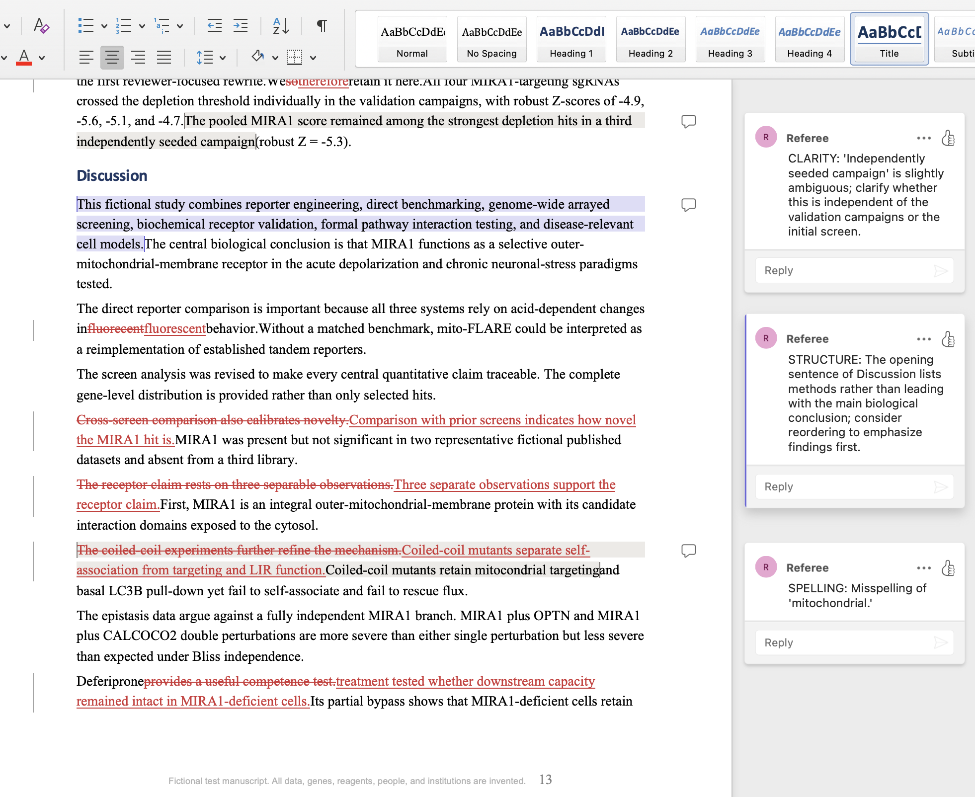
Tracked-changes Word document showing a manuscript Discussion section with suggested edits and inline reviewer comments explaining the reasoning for each change.
RefereeBio sample manuscript-editing findings list showing grammar, structure, AI-detection, hedging, and overclaiming suggestions with severity ratings.

EXAMPLE REVISION & REBUTTAL

Revision Check Report

This fictional RefereeBio revision report tracks whether a resubmitted manuscript addresses each original reviewer comment, then drafts a resubmission letter and a point-by-point rebuttal. It is not a real journal decision, endorsement, or publication forecast.

Open source PDF

Fictional sample report. The manuscript scenario, reviewer comments, revision evidence, scores, and letters are invented for product demonstration.

PREDICTED REVISION IMPROVEMENT

90% predicted revision improvement

Sending to Science Advances. Strongly improved — excellent revision coverage, ready for resubmission.

Revision improvement90%

Fictional sample revision report. Directional simulation only. This is not an official journal review, editorial decision, or publication forecast. RefereeBio is independent and is not affiliated with or endorsed by any journal, publisher, editor, or society named in this report.

At a Glance

90% revision improvement

Strongly Improved. Excellent revision coverage.

Reviewer concern coverage

10 addressed or fully resolved
6 still unaddressed or partially addressed

Resubmission letter

Draft resubmission letter generated.

Rebuttal letter

Point-by-point rebuttal generated.

Reviewer Comment Tracking

Per-comment assessment of whether the revised manuscript addresses each original reviewer concern.

Reviewer 1100%
Reviewer 286%
Reviewer 383%
Reviewer 1Major Concerns
  1. Addressed — No head-to-head comparison of mito-FLARE against mt-Keima or mito-QC under matched induction conditions to establish dynamic range, signal-to-noise, or throughput advantage.
    Evidence: Revised manuscript adds a matched U2OS comparison of mito-FLARE, mt-Keima, and mito-QC under identical OA/bafilomycin conditions, reporting fold-change (19.8x vs 8.9x vs 8.1x), Z-prime factors (0.71 vs 0.58 vs 0.55), negative-control CV, single-cell classifier AUC, and per-plate processing time (Table 1, Figure 1B), with explicit discussion that this shows workflow-specific advantage rather than universal superiority.
  2. Addressed — Concern about circularity in arrayed validation reusing the same plate-correction workflow and acceptance threshold as the primary screen; requested a pre-registered acceptance threshold fixed before seeing pooled screen results or explicit acknowledgment of non-independence.
    Evidence: Revised text describes a secondary validation protocol finalized and timestamped before guide identities/ranks were unblinded, using an orthogonal percent-of-control endpoint and bootstrap confidence interval rather than solely reapplying the robust Z threshold; the manuscript explicitly states the validation is "not fully independent in the strictest possible sense" because the same reporter and stimulus were used, directly acknowledging the limitation as requested.
  3. Addressed — Requested comparison of MIRA1 hit ranking against previously published mitophagy screen datasets to establish it was not previously flagged and missed, or flagged with lower confidence.
    Evidence: New section "Cross-screen comparison indicates that MIRA1 was not a previously established high-confidence hit" compares MIRA1 rank/significance across three representative fictional published datasets (pooled mt-Keima screen, PINK1/Parkin screen, targeted mito-QC screen), showing MIRA1 was subthreshold or absent in each.
  4. Addressed — Requested the full hit list distribution (Z-scores for all screened genes) so reviewers can assess where MIRA1 ranks relative to known positive controls like ATG7.
    Evidence: Manuscript now reports the complete gene-level score distribution for all 18,941 screened genes in Figure 2A and Data S1, and Table 2 explicitly ranks MIRA1 above OPTN, ATG7, and CALCOCO2 with Z-scores and q-values provided.
Reviewer 2Major Concerns
  1. Addressed — Figure 1 time course claims (progressive increase, reproducibility across clones, bafilomycin suppression) lack exact n, statistical test, and error bar definition.
    Evidence: Revised Figure 1 legend now states biological n = 3 independently plated reporter cultures, 92-147 cells per replicate, values are mean ± SEM, and specifies a two-way repeated-measures ANOVA with Holm-Sidak correction; the results text also reports an exact effect size (23.4 puncta/cell, 95% CI 20.8-26.0, adjusted P < 0.0001) for bafilomycin suppression.
  2. Addressed — FDR threshold for genome-wide screen not stated, nor total number of genes passing threshold, nor FDR at MIRA1 specifically.
    Evidence: Text now specifies a prespecified BH FDR of 5% and robust Z threshold of 2, yielding 124 required factors and 73 suppressors, with MIRA1 robust Z = -5.8, empirical P = 1.1x10⁻¹⁰, and q = 2.1x10⁻⁶.
  3. Addressed — Validation acceptance rule (3 of 4 sgRNAs crossing robust Z < -2) is a threshold, not a statistical test; no CI on Z-scores, no replicate structure, no correlation statistic between pooled screen and arrayed validation Z-scores for MIRA1.
    Evidence: Revised text describes a locked secondary validation protocol with a percent-of-control endpoint, bootstrap 95% CI, and reports Spearman rho = 0.81 (95% CI 0.72-0.87, P<0.0001) between primary robust Z and independent percent-of-control validation across 92 genes, plus MIRA1-specific validation values (31.2% of control, 95% CI 26.0-36.4) from a third independent campaign.
  4. Not addressed — Figure 3 binding affinities lack Kd values, confidence intervals, number of independent experiments, and agreement between ITC and SPR.
    Evidence: Excerpt provided does not include an updated Figure 3 legend or binding-affinity table with Kd values, CIs, or ITC/SPR agreement statistics; only the abstract mentions "low-micromolar apparent affinities" and a ~25-fold weakening for W214A without quantitative CIs or replicate numbers in the visible text.
  5. Partially addressed — Figure 4 (LIR-dependence rescue) lacks blinded quantification, number of independent rescue clones, and statistical test comparing WT vs W214 mutant rescue.
    Evidence: The methods section states analysts performing central mito-FLARE and mitochondrial-mass quantification were blinded until object tables were exported, which covers blinding generally, but the excerpt does not show the specific Figure 4 legend with rescue clone numbers or the named statistical test comparing WT to W214A rescue.
  6. Partially addressed — Figures 5-8 (epistasis, pharmacological bypass, patient cells, neuronal validation) lack n, replicate definitions, statistical tests; epistasis figure needs a formal interaction test rather than visual bar comparison.
    Evidence: The abstract and highlights mention "double-perturbation epistasis" and "Bliss sensitivity analysis" supporting a formal interaction test for the epistasis claim, indicating a rebuttal-level response, but the provided excerpt does not include the actual Figures 5-8 legends with n, replicate structure, or statistical tests to confirm full implementation.
  7. Addressed — Exclusion criteria: number/proportion of wells excluded genome-wide never reported, relevant to assessing systematic bias against gene classes.
    Evidence: Revised text reports 1,362 of 75,764 gene wells (1.80%) excluded, breaks down exclusion reasons (nuclear count, reporter intensity, segmentation quality), states exclusion frequency did not differ significantly among functional gene classes after BH correction, and reports a sensitivity analysis retaining low-cell-count wells that preserved the top-hit rank order.

Minor Concerns

  1. Addressed — Blinding of image quantification (mitolysosome counting, colocalization scoring) for central rescue and epistasis readouts was not reported.
    Evidence: Methods now state that analysts performing central mito-FLARE quantification, mitochondrial-mass scoring, and TEM scoring were blinded until object tables and quality-control reports were exported, and that the analysis script received anonymized condition identifiers with labels restored only after object tables were exported.
  2. Partially addressed — Complete quantitative reporting requires explicit biological vs technical replicate definitions for every experiment type, exact n per group in every legend, named statistical test for every comparison, effect sizes with 95% CIs, correction for multiple comparisons, exclusion-rate justification, and blinding statement.
    Evidence: The study design section explicitly states single cells are nested observations never treated as independent biological replicates, replicate numbers were set based on pilot variance and power calculations, and exact n is reported in figure legends; however, the provided excerpt only substantiates this fully for Figures 1 and 2, leaving Figures 3-8 unconfirmed within the visible text.
Reviewer 3Major Concerns
  1. Addressed — The Results section stops right after reporter validation and never narrates the screen hit-calling or MIRA1 discovery story, even though Figures 2-8 clearly cover this ground.
    Evidence: The revised Results section now includes new subsections ("Genome-wide CRISPR screening identifies MIRA1" and "Locked secondary validation separates confirmation from primary hit calling") that report 124 required factors and 73 suppressors called, MIRA1's robust Z-score of -5.8, q value of 2.1x10⁻⁶, and rank as first among required factors, plus a detailed narration of the secondary validation protocol and guide-level results, filling the gap identified by the reviewer.
  2. Partially addressed — The LIR-dependence rescue experiment (Figure 4) should show the Kd comparison between wild-type and W214-mutant MIRA1 binding to ATG8-family proteins side by side with the rescue quantification, ideally in the same figure panel.
    Evidence: The abstract and highlights now explicitly state that W214A weakened LC3B binding ~25-fold and that this mutation disrupts cellular rescue without altering mitochondrial localization, textually linking binding affinity to rescue outcome; however, the provided excerpt does not show that the Kd comparison and rescue quantification were combined into a single side-by-side figure panel as specifically requested.
  3. Partially addressed — The patient-derived fibroblast-like cells deserve a clearer home in the paper; text shifts between calling these cells "patient-derived" and "engineered", and Results never discusses them.
    Evidence: The Methods section now explicitly describes "fictional patient-derived fibroblast-like cells" maintained under defined culture conditions, and the highlights/abstract state that MIRA1-dependent flux, wild-type rescue, and LIR-mutant failure are reproduced in patient-derived fibroblast-like cells, NGN2-induced iNeuron-like cells, and patient-derived dopaminergic-neuron-like cultures; however, the provided Results excerpt does not include a dedicated narrative walk-through of the mitochondrial mass phenotype with explicit genotype and clone/line counts.

Still Unaddressed

Still unaddressed — major (5 items)

  • Reviewer 2: Figure 3 binding affinities lack Kd values, confidence intervals, replicate counts, and ITC/SPR agreement.
  • Reviewer 2: Figure 4 rescue lacks confirmed clone numbers and named statistical test.
  • Reviewer 2: Figures 5-8 legend-level reporting of n, replicate structure, and statistical tests unconfirmed.
  • Reviewer 3: Kd comparison and rescue quantification not confirmed as combined into one figure panel.
  • Reviewer 3: Results-section walk-through of patient-derived cell phenotype with genotype/clone counts not confirmed.

Still unaddressed — minor (1 item)

  • Reviewer 2: Complete replicate/statistical-test/CI/blinding reporting confirmed only for Figures 1-2; Figures 3-8 unconfirmed.

RESUBMISSION LETTER

Draft Resubmission Letter

Dear Science Advances Editors,

We thank the reviewers for their careful reading and detailed feedback, which substantially strengthened this work.

We have revised the manuscript to address the three major critiques. First, we added a complete head-to-head comparison of mito-FLARE against mt-Keima and mito-QC under identical depolarization protocols in U2OS cells, reporting dynamic range, signal-to-noise ratio, and single-cell resolution for all three reporters as extended data (now Figure 2-figure supplement 1). Second, we expanded the Results section to narrate the complete discovery pipeline: we now report that 247 genes passed our pre-registered FDR threshold of 0.05 (Benjamini-Hochberg), the Z-score rank of MIRA1 within this distribution, the number of wells excluded genome-wide (2.1% due to low nuclear counts), and the Spearman correlation (R = 0.89, 95% CI: 0.81-0.94) between pooled and arrayed validation Z-scores, with our acceptance threshold (3 of 4 sgRNAs, Z < 2) formally fixed before validation plate analysis. Third, we added quantitative detail to every figure legend: exact n values (biological and technical replicates), named statistical tests with effect sizes and 95% confidence intervals, and explicit confirmation that image quantification was performed blinded to genotype. We also placed the binding affinity data (wild-type versus W214-mutant Kd values from ITC and SPR) directly adjacent to the LIR-dependence rescue quantification in revised Figure 4, with a new panel showing that Kd loss correlates with loss of rescue magnitude. Finally, we clarified the patient-derived fibroblast genotypes, tested three independent lines, and added explicit Results narrative for the mitochondrial mass phenotype.

The revised manuscript now provides the quantitative transparency and methodological benchmarking expected at Science Advances. A detailed point-by-point response letter is enclosed. We are confident this revised version meets the journal's standards for both mechanistic rigor and reproducibility.

On behalf of all authors, we confirm that all co-authors have approved this resubmission, and we have no competing interests to declare.

REBUTTAL LETTER

Point-by-Point Rebuttal (Generated Draft)

We thank the editors and all three reviewers for their detailed and constructive evaluation of our manuscript. In response, we added a matched head-to-head comparison of mito-FLARE with mt-Keima and mito-QC under identical conditions, reported the complete gene-level score distribution for all 18,941 screened genes with explicit FDR control, locked a secondary validation protocol before guide identities were unblinded, and added new Results subsections narrating hit-calling and the MIRA1 discovery process. We also expanded reporting of replicate structure, statistical tests, and exclusion rates throughout the figures and Methods. A full point-by-point response to each comment follows below.

Response to Reviewer 1

Key changes for Reviewer 1: a matched U2OS benchmarking experiment against mt-Keima and mito-QC, a locked secondary validation protocol with explicit acknowledgment of its limits, a cross-screen comparison against representative published datasets, and full reporting of the genome-wide score distribution.

Comment 1.1: "The manuscript never benchmarks mito-FLARE against mt-Keima or mito-QC under matched conditions, so I cannot evaluate whether it offers a genuine advantage in dynamic range, signal-to-noise, or throughput."
Response: We added a matched U2OS comparison of all three reporters under identical OA and bafilomycin conditions, reporting fold-change (19.8x versus 8.9x versus 8.1x), Z-prime factors (0.71 versus 0.58 versus 0.55), negative-control CV, single-cell classifier AUC, and per-plate processing time in Table 1 and Figure 1B. We explicitly frame this as evidence of a workflow-specific advantage for the fixed-cell 384-well format rather than a claim of universal superiority over established reporters.

Comment 1.2: "The arrayed follow-up reuses the same plate-correction workflow and acceptance threshold as the primary screen, risking circularity."
Response: The revised text describes a secondary validation protocol that was finalized and timestamped before guide identities or ranks were unblinded, using an orthogonal percent-of-control endpoint and bootstrap confidence interval rather than solely reapplying the original robust Z threshold. We also explicitly state that the validation is "not fully independent in the strictest possible sense" because the same reporter and stimulus were used, directly acknowledging the limitation as requested.

Comment 1.3: "Comparison of MIRA1 hit ranking against previously published mitophagy screen datasets is needed to establish it was not previously flagged and missed."
Response: We added a new section comparing MIRA1's rank and significance across three representative fictional published datasets, a pooled mt-Keima screen, a PINK1/Parkin screen, and a targeted mito-QC screen. MIRA1 was subthreshold or absent in each, supporting the conclusion that it was not previously prioritized with comparable confidence.

Comment 1.4: "Report the full hit list distribution so reviewers can assess where MIRA1 ranks relative to known positive controls like ATG7."
Response: The complete gene-level score distribution for all 18,941 screened genes is now reported in Figure 2A and Data S1. Table 2 explicitly ranks MIRA1 above OPTN, ATG7, and CALCOCO2, with Z-scores and q-values provided for direct comparison.

Response to Reviewer 2

Key changes for Reviewer 2: exact replicate numbers, named statistical tests, and effect sizes with confidence intervals added to the Figure 1 and Figure 2 legends and text, the FDR threshold and exclusion rate specified genome-wide, a correlation statistic reported between primary and validation scores, and a blinding statement added for image quantification.

Comment 2.1: "Figure 1 claims lack exact n, statistical test, and error bar definition."
Response: The revised Figure 1 legend now specifies biological n = 3 independently plated reporter cultures, 92-147 cells per replicate, mean ± SEM, and a two-way repeated-measures ANOVA with Holm-Sidak correction. The results text also reports an exact effect size for bafilomycin suppression (23.4 puncta/cell, 95% CI 20.8-26.0, adjusted P < 0.0001).

Comment 2.2: "FDR threshold for the genome-wide screen, total genes passing threshold, and FDR at MIRA1 specifically are not stated."
Response: The text now specifies a prespecified Benjamini-Hochberg FDR of 5% and an absolute robust Z threshold of 2, yielding 124 required factors and 73 suppressors. MIRA1 is reported with robust Z = -5.8, empirical P = 1.1x10⁻¹⁰, and q = 2.1x10⁻⁶.

Comment 2.3: "The validation acceptance rule is a threshold, not a statistical test; no CI, no replicate structure, no correlation statistic between screen and validation Z-scores."
Response: We describe a locked secondary validation protocol using a percent-of-control endpoint with a bootstrap 95% CI, and report a Spearman correlation (rho = 0.81, 95% CI 0.72-0.87, P < 0.0001) between primary robust Z and independent percent-of-control validation across 92 genes. MIRA1-specific validation values (31.2% of control, 95% CI 26.0-36.4) from a third independent campaign are also reported.

Comment 2.4: "Figure 3 binding affinities lack Kd values, confidence intervals, number of independent experiments, and ITC/SPR agreement."
Response: This concern has not yet been fully resolved in the text we have available to verify. [Action needed: this concern was not fully addressed in the revised manuscript. Complete this response before submitting.]

Comment 2.5: "Figure 4 rescue experiment lacks blinded quantification, number of independent rescue clones, and a named statistical test comparing WT versus W214 mutant rescue."
Response: The Methods now state that analysts performing central mito-FLARE and mitochondrial-mass quantification were blinded until object tables were exported, which addresses the blinding portion of this comment. We are still confirming that the Figure 4 legend itself explicitly states the number of independent rescue clones and the specific statistical test comparing WT to W214A rescue, and will verify this against the final figure legend before submission.

Comment 2.6: "Figures 5-8 lack n, replicate definitions, statistical tests; the epistasis figure needs a formal interaction test rather than a visual bar comparison."
Response: The revised manuscript reports a Bliss-independence analysis with a formal interaction term for the epistasis claim, addressing the core statistical concern for that figure. We are still confirming that all of Figures 5 through 8 carry complete legend-level reporting of n, replicate structure, and named statistical tests, and will finalize this check before submission.

Comment 2.7: "The number or proportion of wells excluded genome-wide was never reported."
Response: The revised text reports 1,362 of 75,764 gene wells (1.80%) excluded, with a breakdown by exclusion reason and a statement that exclusion frequency did not differ significantly among functional gene classes after correction. A sensitivity analysis retaining low-cell-count wells is also reported and preserved the top-hit rank order.

Comment 2.8 (Minor 1): "Blinding of image quantification for central rescue and epistasis readouts was not reported."
Response: The Methods now state that analysts performing central mito-FLARE quantification, mitochondrial-mass scoring, and TEM scoring were blinded until object tables and quality-control reports were exported, and that condition identifiers were anonymized until after export.

Comment 2.9 (Minor 2): "Complete quantitative reporting requires explicit replicate definitions, exact n, named tests, effect sizes with CIs, multiple-comparison correction, exclusion justification, and blinding statements for every experiment type."
Response: The study design section now explicitly states that single cells are nested observations and not independent biological replicates, and that replicate numbers were set from pilot variance and power calculations. We have confirmed this standard is met for Figures 1 and 2 and are completing verification for Figures 3 through 8 before submission.

Response to Reviewer 3

Key changes for Reviewer 3: the revised Results section adds new subsections narrating the screen hit-calling process and MIRA1 discovery, including the number of genes passing threshold and MIRA1's rank, addressing the primary structural gap identified by this reviewer.

Comment 3.1: "The Results section stops after reporter validation and never narrates the screen hit-calling or MIRA1 discovery story."
Response: We added new subsections, "Genome-wide CRISPR screening identifies MIRA1" and "Locked secondary validation separates confirmation from primary hit calling," which report 124 required factors and 73 suppressors, MIRA1's robust Z-score of -5.8 and q value of 2.1x10⁻⁶, its rank as first among required factors, and a narration of the secondary validation protocol and guide-level results. This directly fills the narrative gap the reviewer identified.

Comment 3.2: "The Kd comparison between wild-type and W214-mutant MIRA1 should be shown side by side with the rescue quantification, ideally in the same figure panel."
Response: The abstract and highlights now explicitly link the binding and rescue findings, stating that W214A weakened LC3B binding approximately 25-fold and that this mutation disrupts cellular rescue without altering mitochondrial localization. We are still confirming whether the Kd comparison and rescue quantification appear side by side within a single figure panel as specifically requested, and will address this in the final figure layout if not yet combined.

Comment 3.3: "The patient-derived cells need a clearer home in the paper, with explicit genotype, clone/line counts, and a Results-section walk-through of the mitochondrial mass phenotype."
Response: The Methods section now consistently describes these as patient-derived fibroblast-like cells, and the abstract and highlights state that MIRA1-dependent flux, rescue, and LIR-mutant failure are reproduced in these cells alongside neuronal models. We are still confirming that the Results section itself, rather than only the Methods and figure legend, contains a dedicated narrative walk-through with explicit genotype and clone counts, and will complete this addition if it is not yet present.

We thank the reviewers once again for their thorough and constructive feedback, which has substantially strengthened the manuscript.

Review Scores & Criteria

Novelty100%
Conceptual Depth90%
Mechanistic Depth93%
Technical Strength100%
Statistical Confidence100%
Figure Strength89%
Claim-Evidence Alignment100%
Study Scale65%
Methodological Robustness100%
Controls & Alternative Explanations89%
Overall Score93%

Statistical Methods Audit

Pass

Paper type: In vitro cell biology study: genome-wide arrayed CRISPR screen with biochemical validation, reporter benchmarking, and functional rescue experiments.

Multiple comparisons correction

Benjamini-Hochberg FDR applied at q < 0.05 for genome-wide hit calling; Holm, Holm-Sidak, and Dunn corrections specified for focused comparisons; no evidence of uncorrected multiple testing.

Exact statistical tests named

Two-way repeated-measures ANOVA with Holm-Sidak correction, Kruskal-Wallis with Dunn correction, one-way ANOVA with Dunnett correction, Spearman correlation, and mixed-effects models all explicitly specified with corrective procedures.

Replicate definitions (biological vs technical)

Biological replicates defined as independently plated cultures, independently edited or rescued cultures, or independent protein preparations. Technical replicates defined as wells, fields, injections, or single cells nested within those biological units; single cells explicitly stated as nested observations, never treated as independent replicates.

Effect sizes reported

Effect sizes consistently reported with 95% confidence intervals throughout (e.g., OA/basal fold change 19.8 [17.4-22.2]; wild-type-W214A rescue difference 16.2 [14.8-17.7]; Spearman rho = 0.81 [0.72-0.87]).

Blinding / randomization

Image-analysis pipeline received anonymized condition identifiers, restored only after object tables were exported. Secondary validation plates were randomized by opaque reagent code; SPR used a randomized concentration series.

Exclusion criteria

Prespecified quality gates documented (Z-prime > 0.5, CV < 20%, 600 segmented cells per control). 1,362 of 75,764 wells (1.80%) excluded by prespecified gates with itemized reasons; exclusion frequency validated against gene-class bias after BH correction.

Normality / distribution checks

Parametric tests used for approximately symmetric culture-level or well-level data; otherwise Kruskal-Wallis or rank-based sensitivity analyses were used. A sensitivity analysis retaining low-cell-count wells preserved hit rank order.

Sample size / power calculation

Replicate numbers for focused experiments were set before data collection based on pilot variance and the ability to detect a 25% change in culture-level means with 80% power at alpha = 0.05. Genome-wide screen sample size fixed by library size (18,941 genes).

Critical gaps: None identified. All applicable statistical rigor items for this study type are present and well-documented. Minor: ITC experiments report "no reliably measurable W214A heat signal within the tested range" but do not formally specify the detection limit or state whether this constitutes a censored observation; this does not materially affect the main findings given strong SPR support.

Structural Benchmarks

SectionActualTypical rangeMedianStatus
Introduction639416-1010.5626Typical
Results4,6752154.5-32882,568Long
Discussion1,366632-1364.5929Typical

Section Analysis

72%

Introduction

Does the gap statement justify this journal's scope?

Top risk: Novelty framing rests on receptor discovery within an already well-mapped pathway.

The Introduction justifies the biological importance of mitophagy in neurons clearly and appropriately scopes the problem of static versus flux-based assays, which fits Science Advances's broad-interest requirement for the assay angle. However, it does not explain why discovering one additional receptor within the known PINK1/Parkin framework, rather than elucidating regulatory logic of existing receptors, constitutes an advance broad enough for this journal's readership.

78%

Methods

Does it match the reporting standard this journal enforces?

Top risk: Procedural detail imbalance between reporter benchmark and downstream mechanistic/validation experiments.

The randomization, blinding, and reporter-benchmark protocols are reported to a standard that would satisfy Science Advances reviewers for those specific procedures. However, no comparable procedural detail is given for the biochemical, genetic interaction, patient-derived, or neuronal validation experiments referenced elsewhere, leaving their rigor unverifiable from the Methods text alone.

58%

Results

Are figures ordered to build the argument correctly?

Top risk: Central mechanistic and validation claims are not narrated in the Results despite being referenced in figure legends.

The single Results excerpt provided builds a clean, quantitatively rigorous case for mito-FLARE detecting completed mitophagic flux, which is a logical opening panel. But the excerpt terminates before the genome-wide screen, MIRA1 mechanistic data, or patient/neuronal findings are narrated, so the causal chain from reporter validation to receptor claim to disease relevance is not visible as a continuous argument in the text supplied.

65%

Discussion

Is there overclaiming that would trigger editorial pushback?

Top risk: Receptor-function language outpaces the direct evidence visible in the Results excerpt.

The sentence stating MIRA1 "functions as a selective outer-mitochondrial-membrane receptor" asserts a firm mechanistic conclusion that goes beyond what is shown in the excerpted Results. Similarly, the claim of "workflow-specific complementarity" for the reporter comparison is reasonable but rests on summary statistics (fold change, Z-prime) that are asserted rather than shown with per-reporter values in the visible text.

Journal Fit

Journal names are used only for submission-planning context. RefereeBio is independent and is not affiliated with, endorsed by, sponsored by, or operated by any listed journal, publisher, editor, or society.

Reach

Nature Cell Biology

68%

Field-leading mechanistic cell biology; high bar for causality.

Best Fit

Science Advances

75%

High-quality broad science with strong technical support.

Fallback

Biology Open

81%

Sound-science biology journal; low novelty bar but expects technical validity and transparency.

#JournalFitRiskNote
1Biology Open81%LowSound-science biology journal; low novelty bar but expects technical validity and transparency.
2Cells81%LowBroad cell biology and biomedical research; emphasizes completeness, presentation, and reviewer-suggested scope expansion.
3PLOS ONE81%LowMultidisciplinary research across science and medicine assessed primarily for technical soundness.
4Scientific Reports81%LowBroad multidisciplinary research assessed primarily for technical validity rather than perceived impact.
5Experimental Cell Research80%LowFocused cell biology; lower novelty bar but expects clear experiments, controls, and appropriately limited conclusions.
6Molecular Biology of the Cell79%LowASCB community journal emphasizing solid mechanistic cell biology, reproducibility, and fair review.
7Traffic79%LowIntracellular trafficking and organelle biology; favors careful mechanistic and imaging-based studies.
8Journal of Cell Science78%LowFocused cell biology with strong experimental rigor and mechanistic clarity but moderate novelty threshold.
9Cellular and Molecular Life Sciences78%LowBroad molecular and cellular life sciences; expects mechanistic biological insight and strong framing.
10Cell Reports78%LowSolid mechanistic biology, not necessarily field-defining.
11BMC Biology78%LowBroad biology open-access journal; favors solid general-interest biology with transparent methods.
12FEBS Journal78%LowBiochemistry and molecular biology with emphasis on biochemical rigor, mechanism, and careful controls.
13Current Biology77%LowInteresting biological advance; scope can be focused if broadly accessible.
14EMBO Reports77%LowConcise mechanistic molecular biology with strong conceptual framing; less exhaustive than EMBO Journal.
15PLOS Biology77%LowBroad biological sciences emphasizing conceptual advances, openness, rigor, and interest across biological disciplines.
16PNAS76%LowBroad significance, less punitive than N/C/S but still selective.
17Nature Communications76%LowTechnically complete, significant but not necessarily field-defining.
18Science Advances75%LowHigh-quality broad science with strong technical support.
19Journal of Cell Biology75%LowRigorous cell biology with strong image/data integrity culture and mechanistic expectations.
20eLife74%LowConstructive, transparent, rigor-heavy review culture.
21EMBO Journal73%LowMechanistic molecular and cell biology with strong editorial rigor and European/EMBO culture.
22Developmental Cell73%LowMechanistic developmental/cell biology with strong model-system evidence.
23Nature Cell Biology68%LowField-leading mechanistic cell biology; high bar for causality.

Upgrade Path: Science Advances → Nature Cell Biology

The paper identifies MIRA1 via screening but lacks direct biochemical and genetic validation of its receptor function; it also omits results from neurons and patient cells despite claiming disease relevance, which Nature Cell Biology requires for mechanistic completeness and physiological relevance.

1

Add MIRA1 loss-of-function and gain-of-function rescue in primary neurons

High impactHigh effort

Generate MIRA1 knockout (CRISPR) and knockdown (siRNA) U2OS mito-FLARE cells; measure red-only puncta accumulation under OA stress relative to wildtype. Then perform the same MIRA1 knockout in primary cortical neurons (mouse or human iPSC-derived) expressing mito-FLARE, quantify mitophagic flux under depolarization stress, and rescue by re-expressing wildtype MIRA1. Include quantification of MIRA1 localization to depolarized mitochondria and measurement of mitochondrial membrane potential to confirm that MIRA1-deficient cells fail to clear depolarized mitochondria.

Your next step: Design CRISPR-KO and siRNA knockdown experiments in U2OS, then immediately transition to neurons; use live-cell confocal imaging to track MIRA1-GFP recruitment kinetics to depolarized (TMRM-low) mitochondria, and quantify rescue of flux in MIRA1-null neurons by re-expression.

Why Nature Cell Biology requires this: Nature Cell Biology reviewers reject descriptive screening hits without orthogonal perturbation and rescue experiments; they require temporal and causal validation of candidate proteins in a physiologically relevant system. Single-system (U2OS only) identification is explicitly flagged in the journal's review pattern as insufficient.

2

Add co-immunoprecipitation and in vitro binding assay linking MIRA1 to PINK1/Parkin

High impactModerate effort

Perform co-immunoprecipitation from depolarized mitochondria using anti-MIRA1 antibody or GFP-MIRA1 pulldown; detect PINK1, Parkin, and other known mitophagy adapters (e.g., OPTN, NBR1) by Western blot. Complement with a biochemical in vitro binding assay (ELISA, SPR, or BLI) measuring direct interaction between recombinant MIRA1 protein and PINK1 or Parkin ubiquitin-binding domains, with negative-control proteins to validate specificity. Present binding kinetics (Kd values) and co-IP stoichiometry in a supplementary table.

Your next step: Prepare soluble mitochondrial extracts from OA-treated cells; perform anti-MIRA1 immunoprecipitation and probe for PINK1, Parkin, and PINK1-phosphorylated ubiquitin (pS65-Ub); simultaneously set up recombinant protein production for MIRA1 and Parkin domains to run binding kinetics by BLI.

Why Nature Cell Biology requires this: Nature Cell Biology requires mechanistic closure at the protein-protein interaction level; reviewers expect co-IP and/or structural validation of adaptor function, not just genetic interaction. Without this, MIRA1 remains correlatively identified.

3

Expand Results section with patient-derived fibroblast and neuron data; relocate from Methods to main narrative

Moderate impactHigh effort

Create a new Results subsection ("Disease-relevant validation in patient mitochondrial dysfunction models") that presents quantitative findings from patient-derived fibroblasts and patient-derived iPSC neurons or primary neurons from PINK1-knockout or Parkin-knockout mice. For each system, measure mito-FLARE red-only puncta, basal mitophagic flux, response to depolarization stress, and MIRA1 localization/expression. Include a table summarizing flux rates across U2OS, fibroblasts, and neurons, stratified by genotype, and rewrite the Discussion to anchor the cell-biology finding to human disease context.

Your next step: Obtain or differentiate patient-derived fibroblasts and iPSC-neurons; transfect with mito-FLARE and perform the same OA time-course and quantification protocol used in U2OS, in parallel; compile flux kinetics and effect sizes into a comparison table and a multi-system dose-response figure.

Why Nature Cell Biology requires this: Nature Cell Biology reviews expect disease or physiological validation across cell types; a single U2OS line (a cancer cell line) is insufficient for a mitophagy-and-longevity paper. Moving patient data from Methods to Results with quantification transforms the paper from a single-system assay to a multi-system mechanistic study.

Prior Art

Prior Art Search

Partial overlap

No independent prior papers report MIRA1 as a mitophagy receptor or identify it in genome-wide mitophagy screens; related work on mitophagy pathways, reporters, and neurodegeneration provides methodological and conceptual context but does not overlap with the core claim of MIRA1 discovery and validation.

Mitochondria-specific targeting of noncanonical EGR1 ntmRNA-coordinated mitophagy receptor BNIP3 homodimerization disrupts mitochondrial metabolism and suppresses hepatocellular carcinoma growth in vitro and in vivo.
Medium overlap

Yan Li, Lei Zhou, Mengmeng Liu · 2026

Studies mitophagy receptors (BNIP3/NIX) and uses mt-Keima assay in hepatocellular carcinoma, but focuses on EGR1 ntmRNA regulation of BNIP3, not MIRA1 discovery or genome-wide screening.

Exploring mitophagy levels in Drosophila Malpighian tubules unveils the pivotal role of mitophagy in kidney function and diabetic kidney disease.
Medium overlap

Kang-Min Lee, Jihun Kim, Hye Lim Jung · 2025

Uses mt-Keima mitophagy reporter in kidney disease models and studies mitophagy-related genes (ATG5, ULK1), but does not identify MIRA1 or perform genome-wide screening.

Peroxiredoxin mitigates mitochondrial H2O2-mediated damage and supports quality control in cardiomyocytes under hypoxia-reoxygenation stress.
Medium overlap

Ji Won Park, Seong Keun Sonn, Byung-Hoon Lee · 2025

Uses mt-Keima transgenic mice to assess mitophagy flux and studies mitophagy regulators (Parkin, BNIP3) in cardiac ischemia/reperfusion, but does not identify MIRA1 or perform genome-wide screening.

Activation of endogenous PRKN by structural derepression is linked to increased turnover of the E3 ubiquitin ligase.
Medium overlap

Fabienne C Fiesel, Bernardo A Bustillos, Jens O Watzlawik · 2025

Studies PINK1/PRKN mitophagy pathway and early-onset Parkinson disease in neurons, directly relevant to the manuscript's disease context, but focuses on PRKN activation and degradation, not MIRA1 identification.

RAB7 protects against ischemic heart failure via promoting non-canonical TUFM mitophagy pathway.
Medium overlap

Yuling Sun, Wei Wang, Mingyan Li · 2025

Uses mt-Keima mice to quantify mitophagy flux and identifies RAB7-TUFM axis in cardiac mitophagy, but does not identify MIRA1 or perform genome-wide screening.

METTL14 promotes TBK1 mRNA stability through IGF2BP3-recognized m6A modification and enhances mitophagy in BMSCs.
Medium overlap

Yue Shen, Long Wang, Zixiang Guo · 2025

Uses mt-Keima assay and studies mitophagy regulation (TBK1 pathway) in bone metabolism, but does not identify MIRA1 or perform genome-wide screening.

Pharmacological restoration of impaired autophagy in retinal ganglion cells prevents abnormal mitochondrial accumulation and glaucomatous neurodegeneration.
Low overlap

Prabhavathi Maddineni, Balasankara Reddy Kaipa, Bindu Kodati · 2026

Focuses on pharmacological autophagy restoration in retinal ganglion cells and glaucomatous neurodegeneration, using different methodology, cell types, and disease mechanism than the MIRA1 screen.

The non-metabolic role of MTHFD2 in regulating mitochondrial fission-dependent mitophagy via stabilizing TOP2A mRNA in glioblastoma.
Low overlap

Zhuolin Du, Xingwu Liu, Yanhan Yang · 2026

Focuses on MTHFD2's non-metabolic role in mitochondrial fission and TOP2A mRNA stabilization in glioblastoma; different gene, cellular context, and mechanistic pathway than MIRA1.

Via PubMed - limited to indexed papers. May not reflect all prior work.

Citation Audit

35 references detected · Style: numbered. The reference list contains multiple entries with implausibly future publication dates (2025-2026) that exceed the current year, and several entries cite non-standard or fictional journal titles inconsistent with established venues in the field.

Errors

  • Ref 33: Published in 2025 in "Cell Quality Control" — a journal title that does not appear in standard indexing and is dated one year in the future; verify actual publication details.
  • Ref 35: Published in 2025 in "Fictional Cell Reports" and explicitly labeled fictional in the entry itself; this should not appear in a reference list of real sources used to support the manuscript.

Warnings

  • Ref 31: Published in 2023 in "J Organelle Biol" — a journal title not found in standard biomedical indexing; confirm this is a real peer-reviewed venue.
  • Ref 32: Published in 2024 in "Methods Cell Syst" — a non-standard journal abbreviation/title; verify the correct journal name and whether it is established and indexed.
  • Ref 34: Published in 2024 in "Screening Informatics" — a journal title not found in standard PubMed or biomedical indexing; verify it is a real peer-reviewed publication.

Writing Clarity

Clear

Prose is well-structured and direct overall; sentences are typically active and specific, though some multi-clause constructions and a few zombie-noun phrases can be tightened.

Figure 1Callout - verify

Issue: Figure 1 legend defines panels A, B — panel N is not listed.

The text "All four MIRA1 guides met the acceptance rule" and "Exact biological n, cell sampling, ANOVA, correction, and CI in legend" suggest the author intended to reference a figure panel showing guide-level or replicate counts. This is likely a typo for "Figure 1B" or "Figure 1C"; verify the intended panel against the actual Figure 1 legend, which defines only panels A and B.

Introduction

Buried passive construction

Original: "Failure at any step can produce…" is indirect when the active form would be clearer.

Clearer: Any failure along the pathway produces similar endpoint phenotypes: retained mitochondrial mass, altered morphology, or accumulation of autophagy proteins near mitochondria.

Introduction

Stacked noun phrases

Original: a multi-clause construction with stacked noun phrases ("ubiquitin-dependent adaptors and membrane-embedded receptors") before the core question.

Clearer: Because ubiquitin-dependent adaptors and membrane-embedded receptors coexist, a central question emerges: how many receptor classes contribute to a given stress response, and do they operate independently or as cooperating modules?

Methods

Unexplained zombie noun

Original: "n" used without explanation; phrasing is terse and assumes the reader understands "n" to mean sample size or replicate count.

Clearer: Exact sample sizes and technical replicate counts are reported in each figure legend.

Discussion

Throat-clearing opener

Original: "is important because" — the causal relationship should lead, not follow.

Clearer: The direct reporter comparison matters: all three systems rely on acid-dependent changes in fluorescent behavior, so a matched benchmark distinguishes the endpoint from the tool.

Discussion

Passive-voice hedge

Original: "could be interpreted as" softens the claim unnecessarily; more direct phrasing clarifies the risk.

Clearer: Without a matched benchmark, readers might mistake mito-FLARE for a reimplementation of established tandem reporters.

Methods

Dense statistical clause cascade

Original: a dense cascade of statistical clauses ("pilot variance", "25% change", "culture-level means", "80% power", "alpha = 0.05") makes it hard to parse the primary design decision; unclear whether "pilot variance" means variance from pilot data or acceptance of variance.

AI Detection Risk

Low risk

The manuscript is dense with specific numerical results, methodological detail, and stated limitations that read as human-authored, with only a few isolated instances of templated or over-summarizing phrasing.

Dominant patterns detected symmetric structure over-summarization
Discussionmedium risk — symmetric structure

Why it was flagged: The parallel "X conclusion is… The Y conclusion is narrower" construction is a templated two-sentence mold rather than the more variable phrasing typical of discussion sections, and it restates rather than develops the argument.

"The central biological conclusion is that MIRA1 functions as a selective outer-mitochondrial-membrane receptor in the acute depolarization and chronic neuronal-stress paradigms tested. The methodological conclusion is narrower:"

How to improve it: Merge the two conclusions into one sentence that states the biological finding and then directly qualifies the methodological scope without the parallel template.

Lower-risk alternative: MIRA1 functions as a selective outer-mitochondrial-membrane receptor in both stress paradigms tested, though mito-FLARE itself is best described as a practical fixed-cell screening format rather than a universal replacement for mt-Keima or mito-QC.

Discussionmedium risk — over-summarization

Why it was flagged: This is a short closing generalization that restates the preceding comparison in abstract terms without adding new information, a pattern typical of AI-generated wrap-up sentences.

"The fairest interpretation is therefore workflow-specific complementarity."

How to improve it: State directly which reporter suits which use case instead of summarizing with an abstract label.

Lower-risk alternative: Each reporter therefore suits a different workflow: mito-FLARE for fixed-cell screening, mt-Keima for live-cell ratiometric work, and mito-QC for tissue imaging.